The Science of Nano-Sized Art
Several beautiful and intriguing images are presented below, graciously made creative commons material by the Nanoscale Informal Science Education Network, a foundation that seeks to improve public engagement in nanoscale science in engineering. Try your hand at guessing what is depicted in each of these images, and when you get stumped, scroll on down to discover the science behind each image, as brought to you by the author of From The Lab Bench. Enjoy!
Tweet
(1) Corralling in The Big One

This image isn’t showing an erupting volcano surrounded by a mountain range… but something far, FAR smaller. Each blue ‘mountainous’ bump is actually an individual iron atom, an atom being the smallest unit structure of an element (like iron). The iron atoms are on a substrate (a surface) of a different element, copper. This structure has come to be known as a quantum coral, created by Don Eigler and colleagues in 1993, who were the first to demonstrate than physical manipulation of individual atoms was possible. Without delving deep into the realms of quantum mechanics, we can describe the wave-like features inside the iron atom ‘corral’ in the following manner: electrons from the copper surface are scattered off of and confined by the ring of iron atoms. It may help to imagine waves in a quiet, circular pond after we throw a rock into its middle. The waves are confined to the pond by land at the shore. Now we can imagine the waves being copper electrons, and the shore being iron atoms. The analogy here is essentially the ‘electron in a box’ description, where copper electrons are trapped inside the ring of iron atoms (in blue). The interference or wave patterns seen inside the quantum corral are the result of the copper electrons (which are classically described as particles) behaving like waves at very small scales. We have a macroscale example of such interference patterns if we throw two rocks into a pond at the same time… we would get interference patterns at the interfaces of the two waves, where some waves are made larger (constructive interference), some are made lower (troughs or valleys caused by destructive interference).

The iron atoms were arranged in a ring via scanning tunneling microscopy (STM). A very sharp (nanoscale to atomic scale) tip can be used to manipulate atoms based on atomic scale forces between the tip and the atom being manipulated.
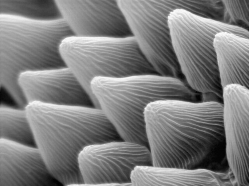
(2) What a Beautiful World

The above image is an electron microscope rendering of wax nanocrystal bundles that help to create what is known as the Lotus effect, the phenomenon that makes the surface of lotus leaves superhydrophobic. The term superhydrophobicity translates into the fact that a water droplet forms a contact angle of greater than 150 degrees with a substrate that has special nanoscale characteristics (i.e. the droplet remains largely spherical even when in physical contact with the substrate). The Lotus effect, which describes the way water beads up and easily rolls off the waxy surface of a leaf, has been used to create superhydrophobic materials like stain-resistant pants and self-cleaning windows. The lotus effect has widespread implications in natural world as well, employed in the insect world to impart self cleaning characteristics to insect legs and wings (Gao 2004, 432, 36). “Water droplets do not stay stably on these surfaces, where they can spontaneously roll off with a slight tremble. During this process, dust particles on the surface are removed.” (Feng 2008).
Dense nanosized features, somewhat like very small whiskers, on the surface of a lotus leaf make it very difficult for water to penetrate into the gaps between these features – this is where surface tension becomes a major factor at the nanoscale, often overpowering the force of gravity which almost always ‘wins’ on the macroscale. In essence, a nano-feature topography affects the way water interacts with the surface. An interface of air remains between gaps between the nano-wax ‘whiskers’ on the surface of the leaf and the layer of water. This means that the wettability of the surface is very low (much of the surface remains dry) and the interactions between the leaf surface and the water are very weak, leading to droplets easily rolling off the leaf or down into a valley at the leaf center.

Water droplets on a hydrophilic surface like normal glass spread out, forming a contact angle of less than 90 degrees. This high wettability is due to absence of the nanoscale features present on the waxy leaf, in other words the surface of the glass is relatively smooth and the water is able to impregnate the entire surface.

An interesting example of nanoscale feature impact on wettability is the rose petal. The rose petal also has very small features on its surface, although these act to a different effect than the waxy nanocrystals on the lotus leaf surface. A rose petal has very small (micro-scale) bumps or micropapillae in between which water can penetrate, allowing a water droplet to form relatively strong interactions with the rose petal surface. However, each micropapillae is covered with even small nanoscale grooves (nanofolds which measure around 760nm), which are too small for water to easily penetrate, similar to the locus effect, creating a superhydrophobic state. The result is a superhydrophobic surface with high adhesive forces (Feng 2008), demonstrated by the fact that a water droplet, although it beads up on the petal surface, will not roll off with ease. In fact, even if we turn the petal upside down, a small water droplet will remain stuck in place on the petal surface.  This superhydrophobic yet adhesive effect is known as the Cassie impregnating wetting state. Another interesting point is that larger water droplets, for example in a hard rain, will side with gravity over the micropapillae-induced adhesive forces, causing the larger drops to roll off the petals and protecting the flower from water-borne damage. It is the unique combination of micro- (10^-6 meters) and nano- (10^-9 meters) structures on the surface of the petal that create the special way that water interacts with the surface. But why would the flower have developed such nanoscale traits? Dr. Feng theorizes that the “petal effect” may “help flowers attract pollinators” by maintaining attractive (to insects) dewdrops on the petal surface.
Gao, X.; Jiang, L. Nature 2004, 432, 36. – Biophysics: water-repellent legs of water striders.
Langmuir 2008, 24, 4114-4119 – Petal Effect: A Superhydrophobic State with High Adhesive Force
Rose Image – Flickr by tanakawho
Nasturtium Leaf Images: A. Otten and S. Herminghaus, Göttingen, Germany, A.Marshall, Stanford University, A.Snyder, Exploratorium
(3) My, What Long Hair you Have…

This image shows a nanowire placed against a human hair. The nanowire (~100nm) measures at least 1000x times less that the average human hair (~100um). Nanowires may have applications in nanocircuitry – making possible smaller and more efficient electronic circuits, and nanophotonics – using the special properties of VERY small materials to guide light with conduits much smaller than the actual wavelength of light, thus overcoming limitations present in, for example, fiber optics. Optical fibers work through total internal reflection, keeping a beam of light completely within the fiber by reflecting the bouncing waves off of its walls. If the diameter of the fiber approaches the wavelength of light (~400nm for blue light) the beam will be able to escape the confines of the fiber walls, making the transfer of light inefficient.
Nanoscale materials on the other hand, especially those composed of metals like silver, have special properties which allow them to produce enhancements in the electric field near the material (a nanoparticle or a nanowire). These near fields around the nanomaterial act to concentrate light to smaller dimensions than the actual wavelength of the light itself! Light can even be converted to surface plasmons (special electron ‘cloud’ oscillations in the material) at one end of a silver nanorod, and converted back to light at the opposite end. This transmission of light is can be, theoretically and increasingly in practice, very efficient. The transmission of light through arrays of nanoscale-sized holes is the principle behind photonic crystals such as the one picture below, which can be used to move light in very precise patterns.

E. Hutter et al. Exploitation of Localized Surface Plasmon Resonance. Adv. Mater. 2004, 16(19)
SEM Image of Nanowire and Human Hair – E Majur, Harvard University
(4) Zoom Zoom Zoom

This is a scanning tunneling microscope image of platinum atoms, arranged in their crystal structure as closely packed hexagonal layers. In this configuration, platinum is very dense, as each atom has the maximum number of directly-touching neighbors. If you have seen a display of oranges forming a pyramid at the supermarket, they were probably placed into closely packed hexagonal layers.

Platinum has applications as a catalyst in automobile catalytic converters and other chemical processing reactions. Efforts are ongoing, for example in the laboratory of Dr. Younan Xia at Washington University in St, Louis, to create more efficient catalytic converters with the use of platinum nanoparticles, which have a higher surface area per unit mass than bulk platinum material.
(5) Achoo!!!

With scanning electron microscopy, we can visualize materials which are much smaller than the wavelength of light (which is a limitation of optical microscopy). This is because instead of using light (photons) to make the image, we use electrons, which can be used to ‘see’ particles as small as 1-5nm. An electron beam is of higher energy than a beam of light, which means the electron beam has a shorter wavelength. The longer wavelength of a light beam and the phenomenon of diffraction (a light beam spreads out when passing through a small hole because photons have wave-light behavior) are what makes optical microscopy only useful for looking at materials larger than 200nm. Diffraction (the spreading out of the wave) means that when we focus light through a small hole in a microscope, the resolution we can achieve is limited because our light beam is not infinitely narrow. In general terms, we can only see things as small as our detection beam is narrow.

The pollen particles shown in the image above are larger than typical nanomaterials, measuring between 6-8 micrometers (um). A micrometer is 10^-6 meters. For comparison, a human hair is typically 50-100 um in diameter. Nanoparticles as typically classified are between 1-100 nanometers, or 1 billionth of a meter. SEM Image of Platinum Atoms – D.Eigler, IBM Almaden Research Center
![]()
Heller, E., Crommie, M., Lutz, C., & Eigler, D. (1994). Scattering and absorption of surface electron waves in quantum corrals Nature, 369 (6480), 464-466 DOI: 10.1038/369464a0
Feng, L., Zhang, Y., Xi, J., Zhu, Y., Wang, N., Xia, F., & Jiang, L. (2008). Petal Effect: A Superhydrophobic State with High Adhesive Force Langmuir, 24 (8), 4114-4119 DOI: 10.1021/la703821h